
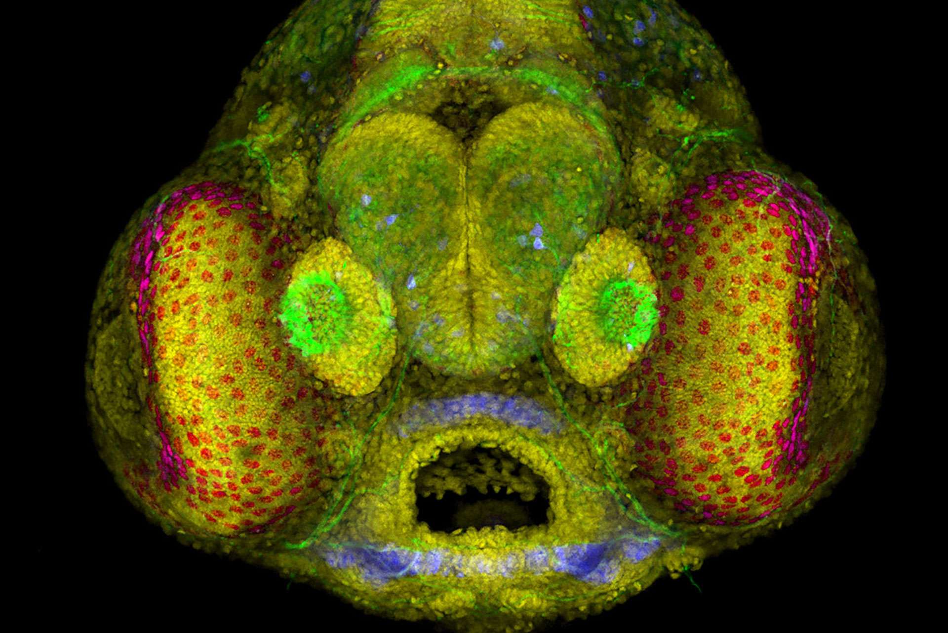
Super-resolution microscopic photography brings never-before-seen images to public view.
Discovering magnificence in photographs so detailed, we are swayed to think the subjects are otherworldly. Microscopic wonders abound all around us, dazzling us with their surreal beauty when captured on film. They appear as extraterrestrial landscapes, fantastical creatures, intricate patterns, and strange structures. What the public cannot see with the naked eye, individuals using super-resolution photomicrography are bringing to light.
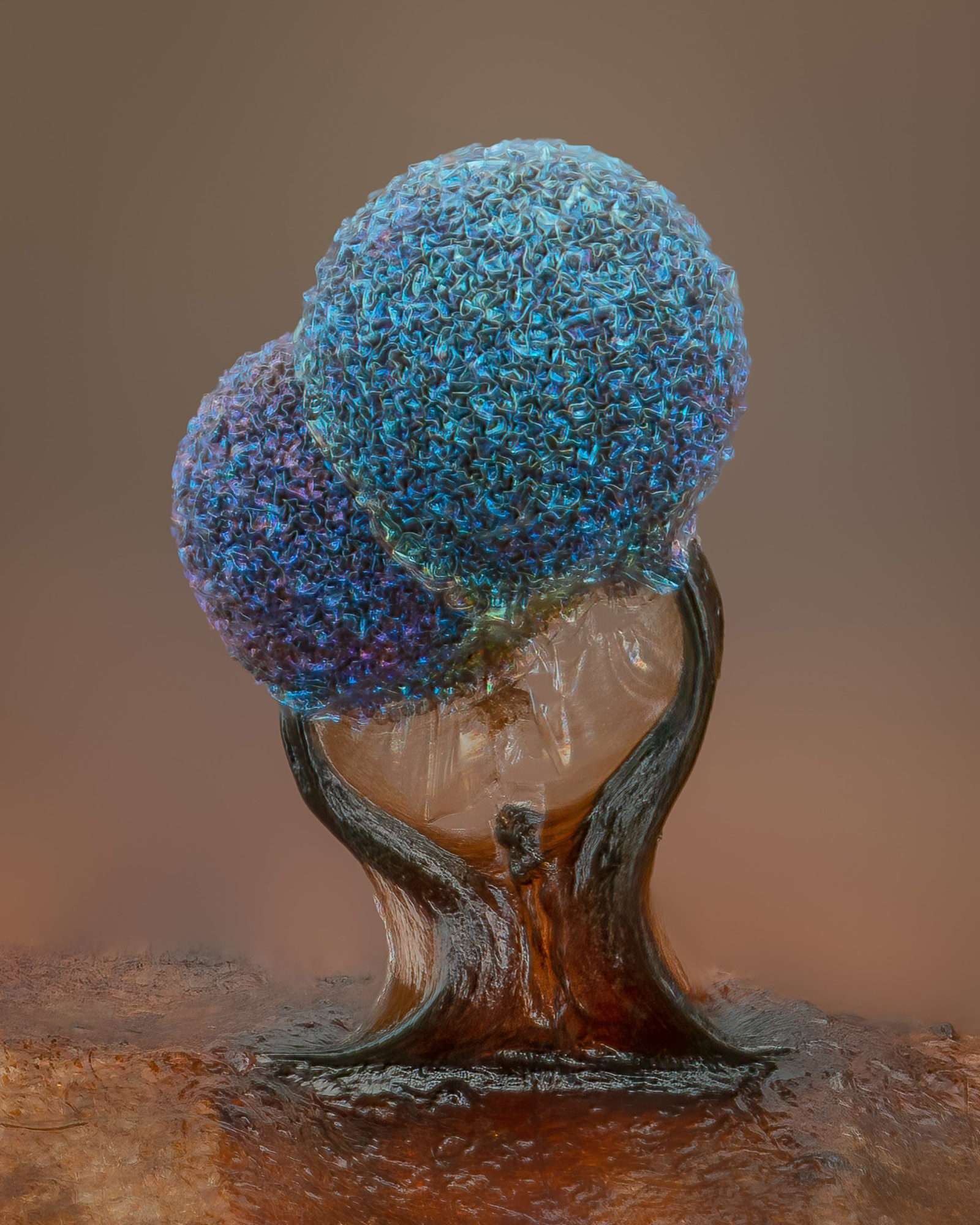
Fifth Place: Slime mold (Lamproderma). Alison Pollack
Recently, Nikon Instruments announced the winners of the forty-eighth annual Nikon Small World Photomicrography Competition. “Each year, Nikon Small World receives an array of microscopic images that exhibit exemplary scientific technique and artistry. This year was no exception,” said Eric Flem, Communications and CRM Manager at Nikon Instruments. “At the intersection of art and science, this year’s competition highlights stunning imagery from scientists, artists, and photomicrographers of all experience levels and backgrounds from across the globe.”
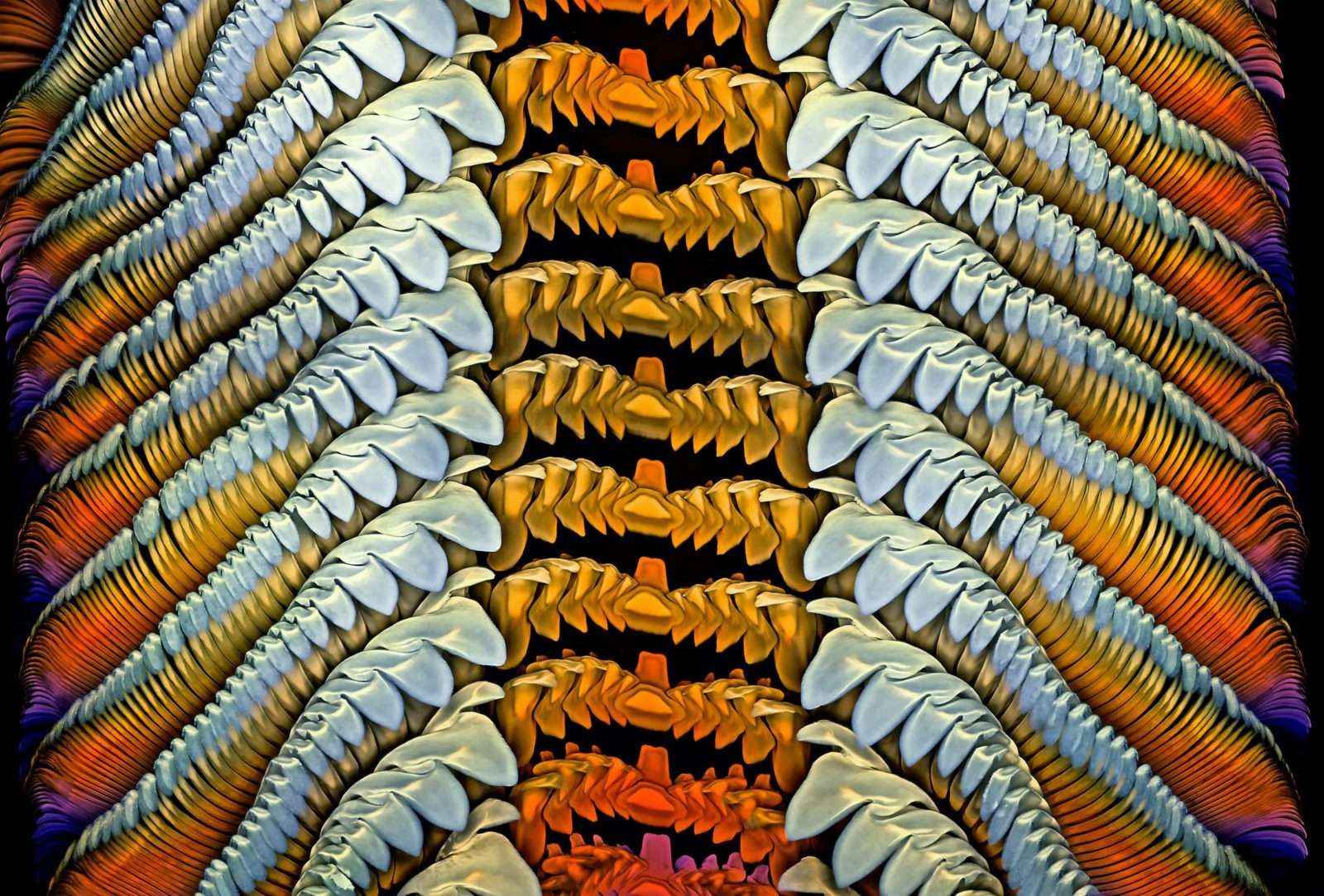
Honorable Mention: Radula (rasping tongue) of a marine snail (Turbinidae family). Dr. Igor Siwanowicz
First Place Winner
The top prize was awarded to Grigorii Timin, supervised by Dr. Michel Milinkovitch at the University of Geneva, Department of Genetics and Evolution. The winning entry is a remarkable image of an embryonic hand of a Madagascar giant day gecko. Artfully rendered and technically immaculate, Timan used high-resolution microscopy and image-stitching to masterfully capture this specimen of the species Phelsuma grandis day gecko.
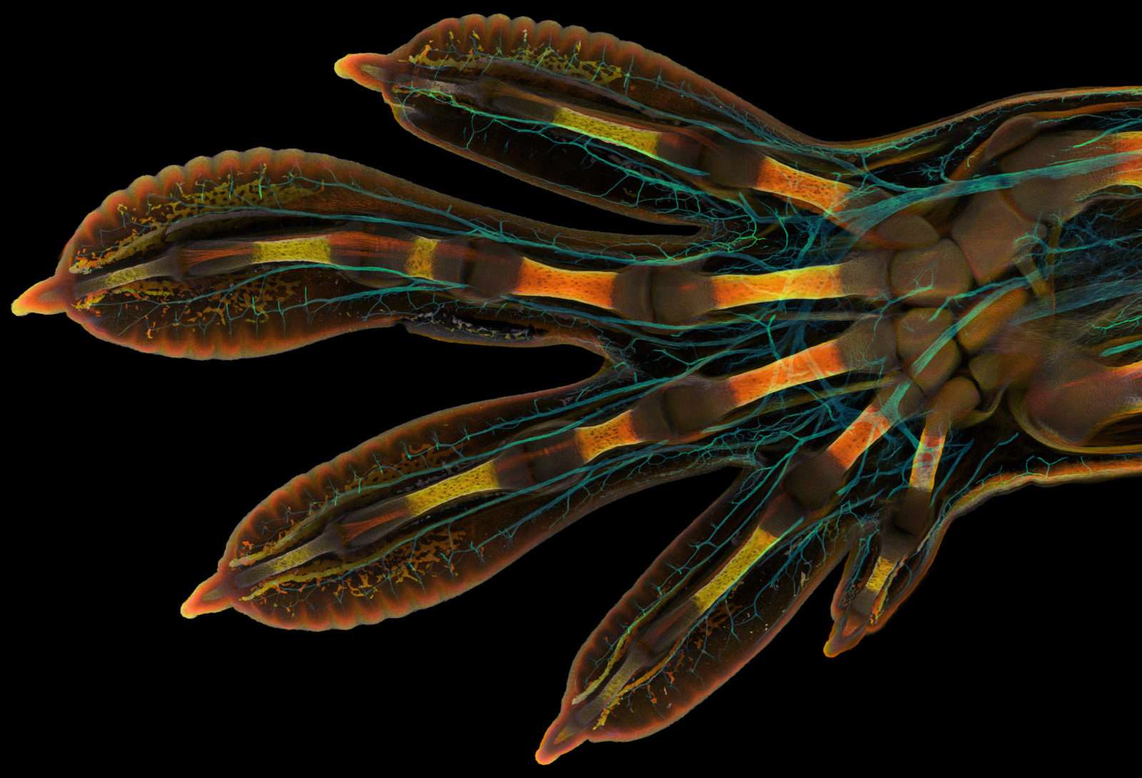
First Place: Embryonic hand of a Madagascar giant day gecko (Phelsuma grandis). Grigorii Timin, Dr. Michel Milinkovitch
Timin painstakingly image-stitched hundreds of images to create this final image. Timin captured the entire embryonic hand using a confocal microscope with whole-mount fluorescent staining and tissue clearing. “This embryonic hand is about 3 mm (0.12 in) in length, which is a huge sample for high-resolution microscopy,” said Timin. “The scan consists of 300 tiles, each containing about 250 optical sections, resulting in more than two days of acquisition and approximately 200 GB of data.”
The visually stunning result reveals the gecko’s hidden beauty and complexity. Nerves were highlighted in cyan color, while the skin, bones, tendons, ligaments, and blood cells were in a range of warmer colors. “This particular image is beautiful and informative, as an overview and also when you magnify it in a certain region, shedding light on how the structures are organized on a cellular level,” said Timin. He went on to say, “The Nikon Small World Competition is a great opportunity to share how impressive nature is on a microscopic level, not only within a scientific community but also with the general public.”
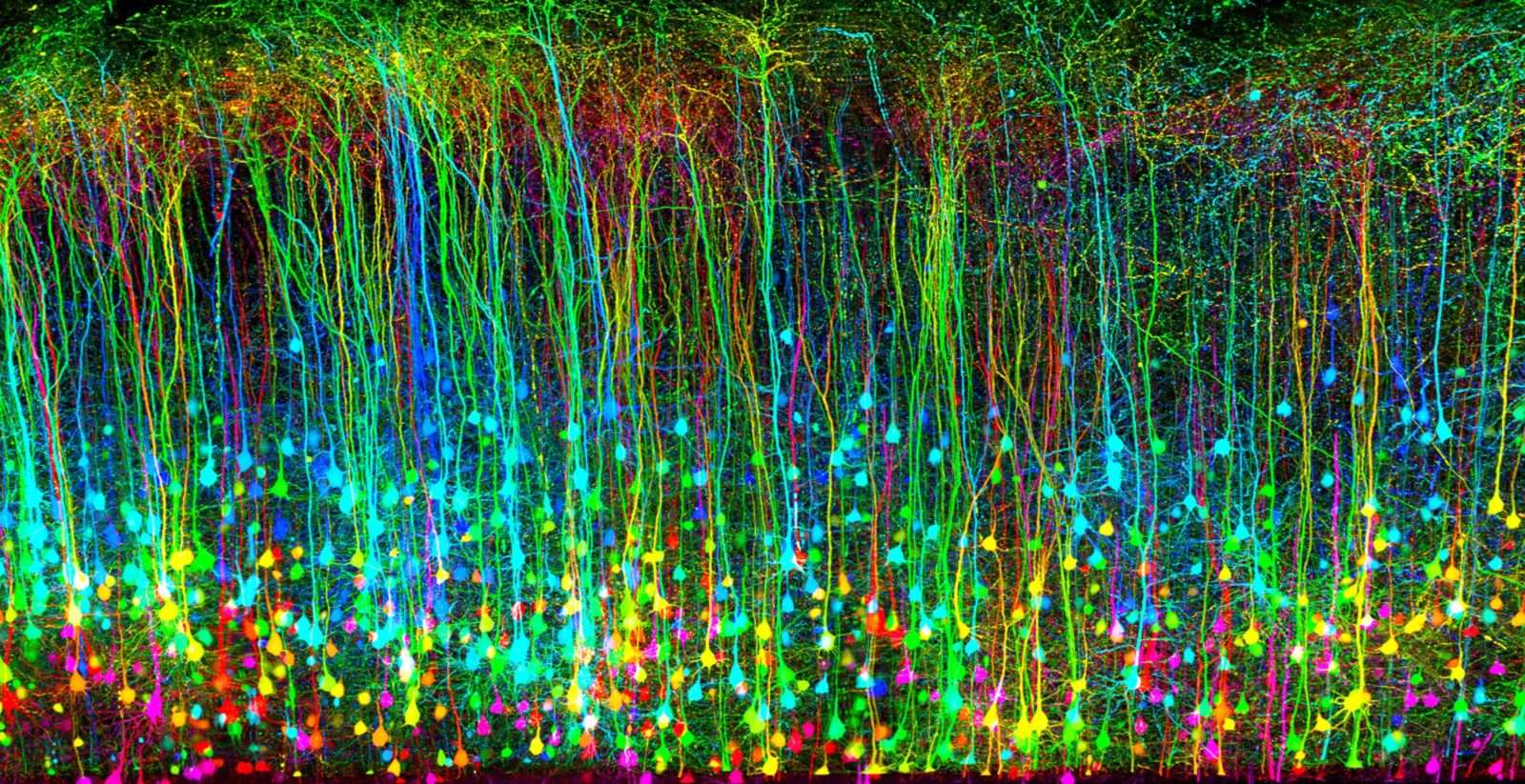
Image of Distinction: Murine sensory-motor cortex following mild traumatic brain injury in a transgenic mouse (expressing Thy1-GFP). Dr. Andrea Tedeschi
Other Nikon Small World Finalists
Second place was awarded to Dr. Caleb Dawson for his image of breast tissue showing contractile myoepithelial cells wrapped around milk-producing alveoli. Taking a week to process, the myoepithelial cells were stained with multiple rounds of fluorescent dyes and captured with a confocal microscope. Dawson is affiliated with WEHI, The Walter and Eliza Hall Institute of Medical Research, Department of Immunology in Melbourne, Victoria, Australia.
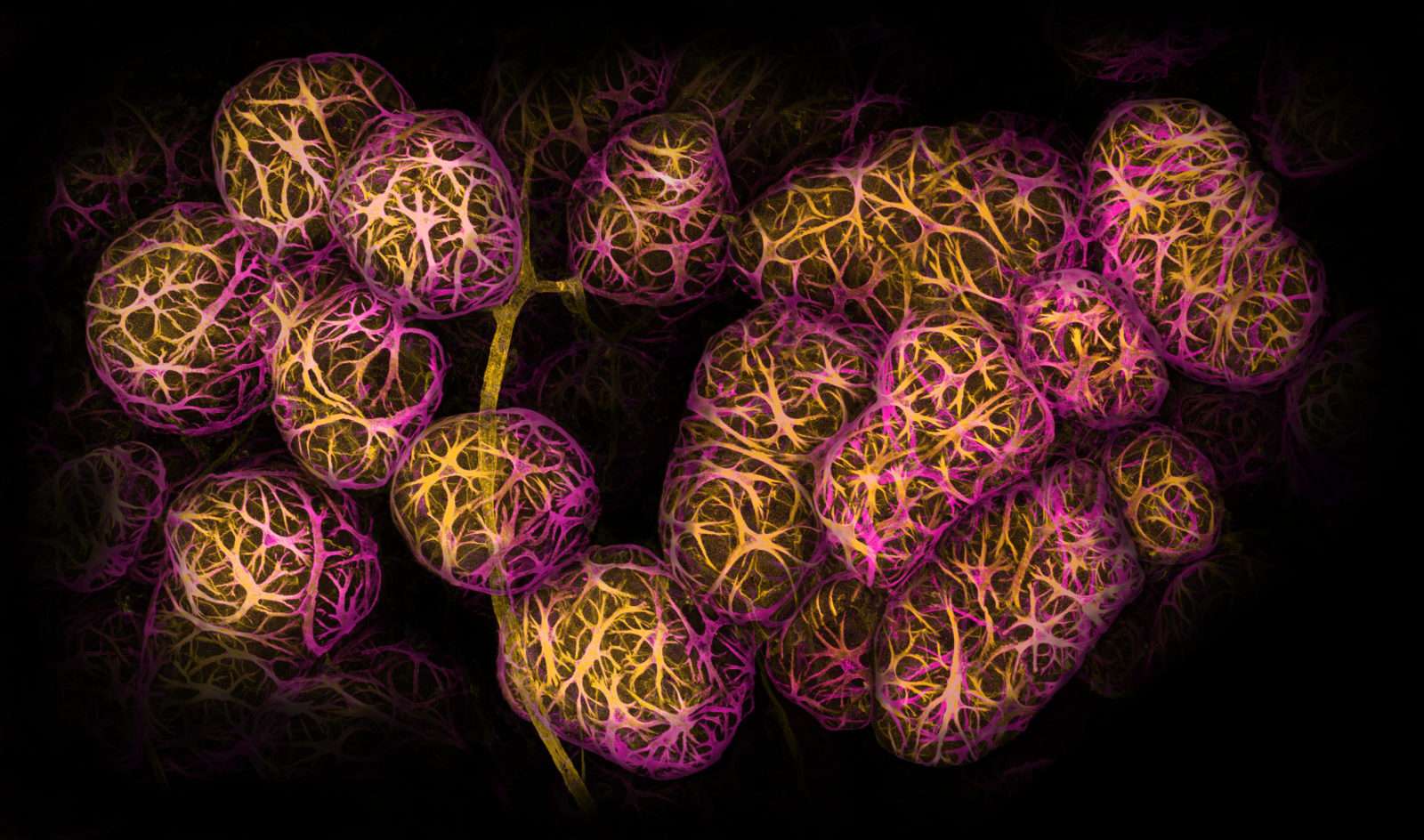
Second Place: Breast tissue showing contractile myoepithelial cells wrapped around milk-producing alveoli. Caleb Dawson
The third place was captured by Satu Paavonsalo and Dr. Sinem Karaman for their image of blood vessel networks in the intestine of an adult mouse. Their Affiliation is with the University of Helsinki, Individualized Drug Therapy Research Program, Faculty of Medicine, Helsinki, Finland.
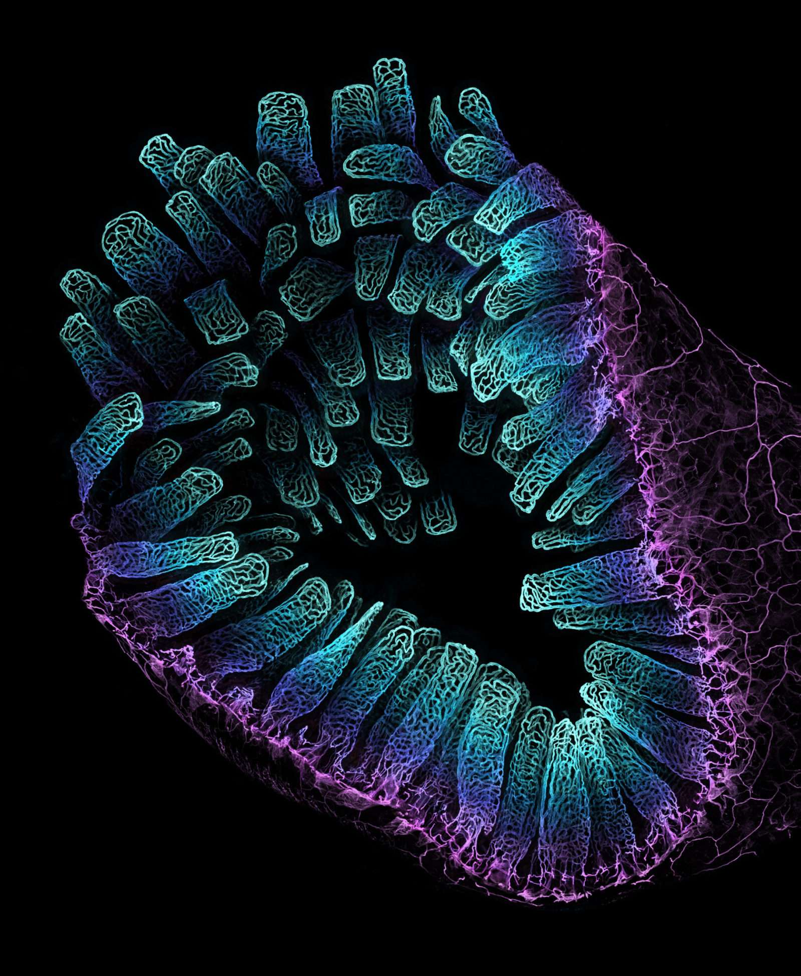
Third Place: Blood vessel networks in the intestine of an adult mouse. Satu Paavonsalo, Dr. Sinem Karaman
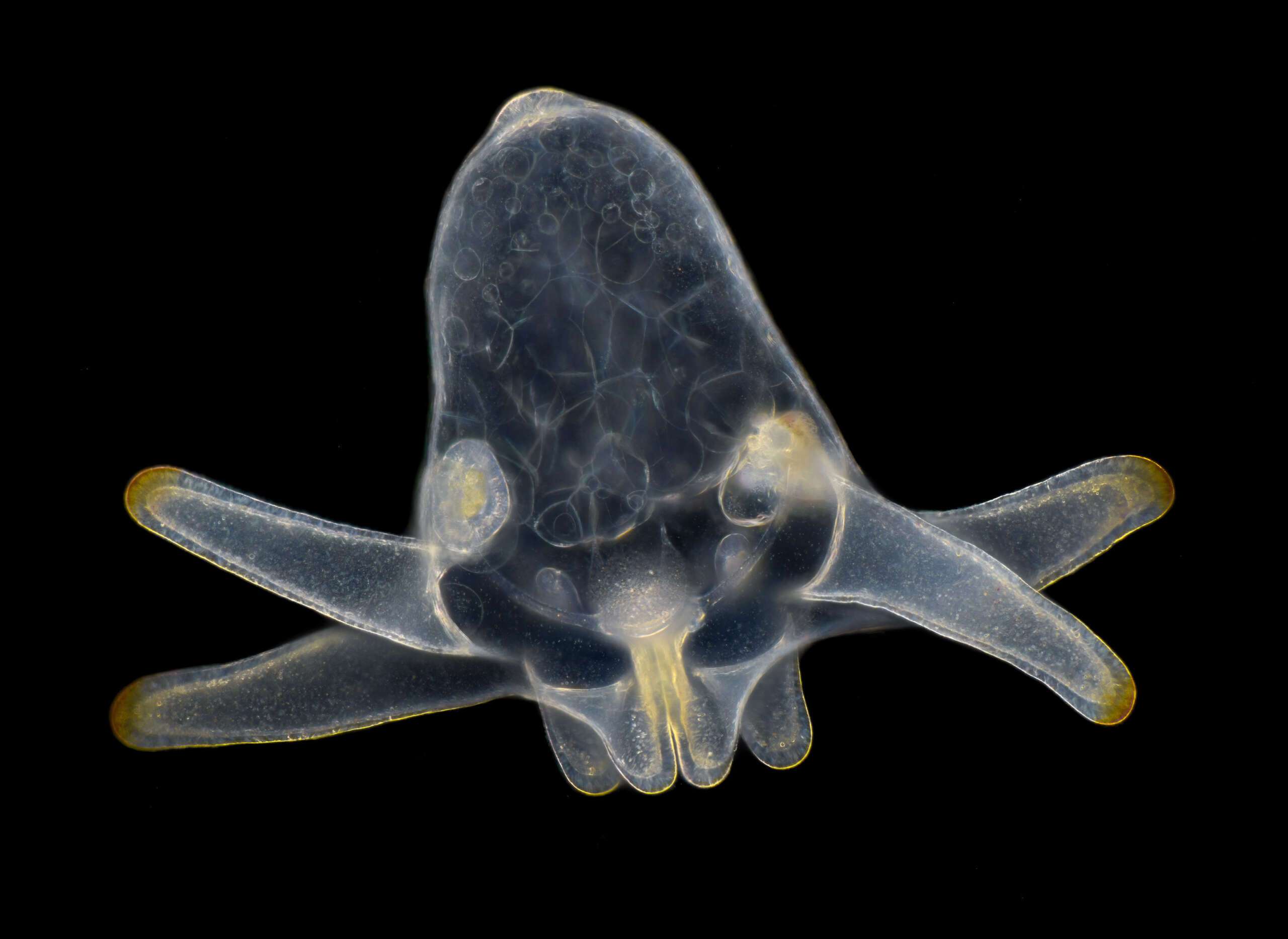
Honorable Mention: Larva of an anemone, found in marine plankton. Wim van Egmond
Nikon Small World gave recognition to 89 photos out of thousands of entries, rounding out the Top 20, Honorable Mentions, and Images of Distinction. Be sure to view the full gallery of Nikon Small World Photomicrography winning images and be dazzled by a miniature world few have ever seen.
Main image award – Image of Distinction: Zebrafish (Danio rerio) embryo head 72 hours after fertilization. Layra G. Cintrón-Rivera.
Contributed by Throomers.